


Innovative Imaging Captures Life in Motion: Nikon’s Small World in Motion Competition Highlights Breakthroughs
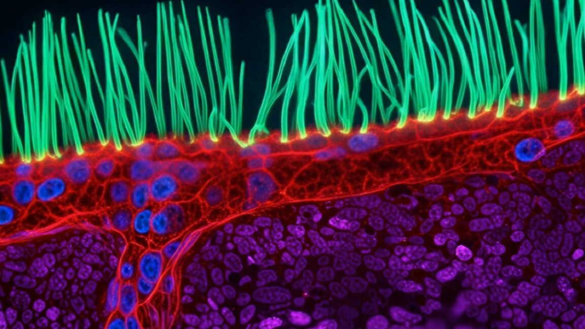
In a remarkable display of technological advancement, the annual Nikon Small World in Motion competition has recognized exceptional talent in the field of microscopy. This year’s winner, Xu, an optical engineer, has pioneered a technique that allows for real-time observation of cellular structures, focusing particularly on the movement of cilia—tiny hair-like projections that play a crucial role in various biological processes.
The ability to visualize cilia in action is vital for understanding diseases tied to their dysfunction. Traditional imaging methods often falter due to the rapid movement and microscopic size of these structures as well as the requirements for capturing them in a living state. Xu’s innovative approach utilizes diffractive super-resolution microscopy, a technique that harnesses multiple light waves and a digital micromirror to finely control light. This process minimizes exposure to the living sample while still achieving high-resolution images of cilia movements.
“To grasp the essence of this disease, observing around ten seconds of motion is crucial—not merely a still image,” Xu articulated. “When viewers witness cilia in motion, it becomes apparent that something intricate and significant is occurring within us. I believe this competition elevates microscopy into a universal language.”
Other impressive contenders demonstrated their work as well. Nguyen Nam Nhat from Vietnam secured second place with a captivating video featuring a tiny roundworm and a single-celled organism. Benedikt Pleyer from Bavaria, Germany, came in third with his visually striking footage of jellyfish larvae observed in water droplets. In fourth place, Andrew Moore from the Howard Hughes Medical Institute in the US presented synchronized cell division in neighboring cells, while Patrick Hickey of Hypha Research Ltd. in Edinburgh claimed fifth place with a video showcasing the dynamics of mitochondria and chloroplasts within Turtle Vine leaf cells.
These winning entries, along with several honorable mentions, can be explored in detail on the Nikon Small World website.
Editor’s Take
This competition not only highlights significant advancements in imaging technology but also emphasizes the importance of visualizing dynamic biological processes. As researchers and developers gain access to such sophisticated techniques, we may see transformative impacts on diagnostics and therapeutic approaches within the life sciences sector. Moreover, the ability to observe cellular functions in real-time fosters a broader understanding of complex biological systems, paving the way for future innovations.
Source: arstechnica.com